
Prosopagnosia: When Faces Refuse to Stick
Published:
There is a particular kind of disorientation in not recognizing a face you’ve seen a hundred times: a colleague, a neighbor, sometimes even your own reflection in a photograph. This is prosopagnosia, or face blindness, the inability to recognize faces despite otherwise normal vision and intact intelligence. The name comes from the Greek prosopon (face) and agnosia (not knowing), and it was first described by the neurologist Joachim Bodamer in 1947.
What makes prosopagnosia so striking is how selective it is. People with the condition can usually still read expressions, judge age or gender, and describe a face feature by feature: the shape of the nose, the color of the eyes. What breaks down is the ability to bind those features into a single, familiar identity. Some people compensate by learning to recognize others through voice, gait, hairstyle, or a distinctive piece of clothing. Oliver Sacks, who wrote about the condition in The Mind’s Eye, disclosed late in life that he had lived with it himself, sometimes failing to recognize his own face in a mirror.
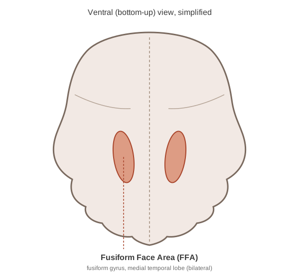
There are two broad forms. Acquired prosopagnosia follows damage to the brain, most often a lesion to the fusiform gyrus on the ventral surface of the temporal lobe. This is the same region that houses the fusiform face area (FFA), a patch of cortex that responds preferentially to faces over almost any other visual category. Developmental prosopagnosia, by contrast, appears without any obvious brain injury. The deficit seems to be present from early in life, as if face-processing circuitry never quite specialized the way it does in most people. Estimates suggest it may affect somewhere around 2% of the population, most of whom never receive a formal diagnosis. They simply think of themselves as “bad with faces.”
What makes the developmental form especially striking is that faces are, by most accounts, the first visual category we ever learn to recognize (Morton & Johnson, 1991). Newborns just minutes old, averaging about nine minutes in the original study, will already track a face-like pattern of three blobs (two “eyes” and a “mouth”) further than a scrambled version of the same blobs (Goren, Sarty, & Wu, 1975), well before they can recognize much else about the visual world. This early bias is sometimes attributed to a subcortical mechanism, Mark Johnson’s “Conspec,” that orients infants toward faces before the cortex has had any real time to learn from experience. It’s thought to bootstrap the cortical face system into existence over the following months. Developmental prosopagnosia is, in that light, less a failure to learn faces and more a case where this head-start specialization never quite consolidates into a working system: a foundational scaffold that in most of us gets built before we can even remember it.
There is no cure for prosopagnosia. A 2014 review of rehabilitation attempts, covering everything from feature-based coaching to perceptual retraining, concluded that no intervention reliably restores normal face recognition, in either the acquired or developmental form (Bate & Bennetts, 2014). What exists instead sits somewhere between management and modest improvement. Most clinical guidance is compensatory: deliberately building the habit of tracking voice, gait, hairstyle, or context instead of the face itself, a workaround rather than a fix, and one that has to be relearned for every new person rather than transferring automatically. What is more interesting, in the spirit of The Brain That Changes Itself, is a smaller body of work asking whether the face system can be retrained rather than just worked around. DeGutis, Cohan, and Nakayama (2014) ran patients with developmental prosopagnosia through weeks of computerized holistic face-processing exercises and found measurable gains on standard face memory tests, in some patients persisting for a year afterward. The effect is real but modest: training tends to improve performance on the specific tasks practiced without fully normalizing everyday recognition, and it does not work for everyone. It is less a dramatic rewiring than the same story told throughout the neuroplasticity literature: the adult brain can shift its tuning with the right repeated input, but slowly, unevenly, and only so far.
A toy simulation: lesioning a face-selective unit
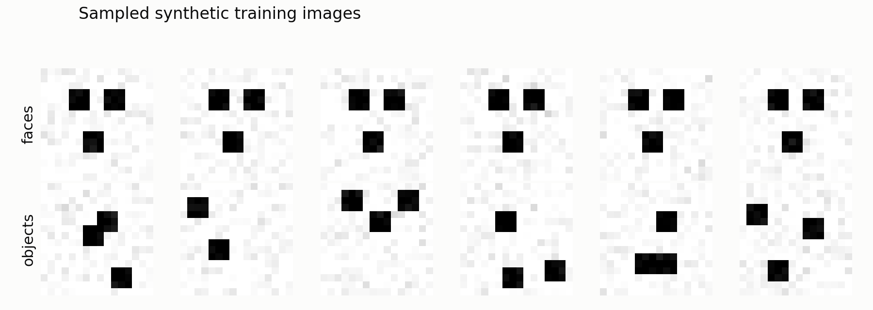
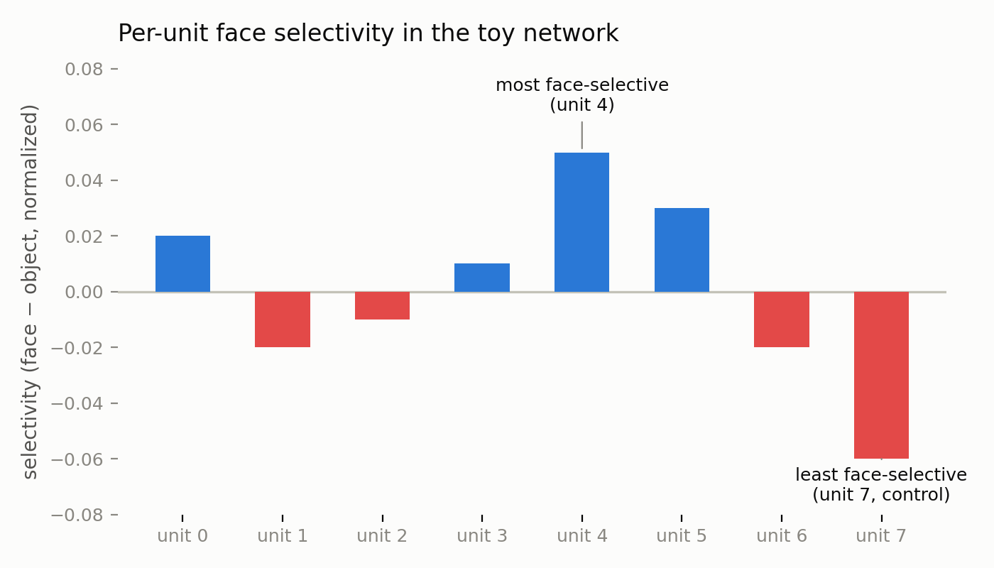
Acquired prosopagnosia is, computationally, a lesion story: damage one patch of cortex and one category of recognition breaks while the rest of vision stays intact. That double dissociation is easy to caricature with a tiny convolutional network. I trained an 8-unit CNN to separate two synthetic categories: “faces” (a fixed eyes/mouth triplet of blobs, always in the same three positions, plus noise) and “objects” (a triplet of blobs at random positions). Then, for each unit, I computed a face-selectivity index (mean activation on faces minus mean activation on objects, normalized), identified the most face-selective unit, zeroed only that unit’s weights, and re-measured accuracy on faces versus objects. As a control, I did the same thing to the least face-selective unit instead.
import numpy as np
import torch
import torch.nn as nn
import torch.optim as optim
import torch.nn.functional as F

torch.manual_seed(0)
np.random.seed(0)


def make_dataset(n=2000, size=16):
    """Synthetic 'faces' (fixed eyes/mouth triplet) vs. 'objects' (randomly placed blob triplets)."""
    X = np.zeros((n, 1, size, size), dtype=np.float32)
    y = np.zeros(n, dtype=np.int64)
    for i in range(n):
        img = np.zeros((size, size), dtype=np.float32)
        if i % 2 == 0:
            for (r, c) in [(4, 5), (4, 10), (10, 7)]:
                img[r - 1:r + 2, c - 1:c + 2] = 1.0
            y[i] = 1
        else:
            for _ in range(3):
                r, c = np.random.randint(2, size - 2, 2)
                img[r - 1:r + 2, c - 1:c + 2] = 1.0
            y[i] = 0
        img += np.random.normal(0, 0.05, img.shape).astype(np.float32)
        X[i, 0] = img
    return torch.tensor(X), torch.tensor(y)


class TinyFaceNet(nn.Module):
    def __init__(self, n_units=8):
        super().__init__()
        self.conv = nn.Conv2d(1, n_units, 3, padding=1)
        self.pool = nn.AdaptiveAvgPool2d(1)
        self.fc = nn.Linear(n_units, 2)

    def forward(self, x):
        h = torch.relu(self.conv(x))
        pooled = self.pool(h).flatten(1)
        return self.fc(pooled), pooled
Sampled examples from that generator, faces on top and objects below:
Training for 30 epochs on 2,000 synthetic images and evaluating on 400 held-out ones gives a per-unit selectivity profile, with one unit standing out as the network’s de facto “FFA”:
Zeroing that single unit, versus zeroing the least face-selective one as a control, produces a sharply category-specific effect:
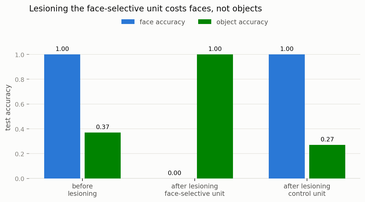
Knocking out unit 4 collapses face accuracy to zero while object accuracy is untouched (in fact it rises, because the network’s decision rule collapses to “always guess object” once its one informative face channel is gone). Knocking out the control unit barely moves either number. It’s a toy in every sense: eight units, one linear readout, a global-average-pool that throws away spatial layout, and a task simple enough that a single filter can carry the whole face signal. Real cortex is nowhere near this clean, and real lesions rarely hit exactly one functional unit. But the qualitative shape, a highly selective unit whose removal costs one category and essentially nothing else, is the same shape researchers have started finding in much larger, more realistic CNNs trained on natural images, where face-selective units emerge unsupervised and behave, on ablation, remarkably like a model FFA (Ratan Murty et al., 2021; Baek et al., 2021).
I find prosopagnosia fascinating for the same reason I find the rest of face perception fascinating. It’s a case study in how much specialized machinery the brain devotes to something that feels effortless. Faces are processed holistically, as a configuration rather than a sum of parts, which is part of why an inverted photo of a face is so much harder to recognize than an inverted photo of almost anything else (the well-known “Thatcher effect” trades on exactly this). Losing that holistic binding, whether through injury or through atypical development, doesn’t erase vision. It removes one very specific, very human shortcut for turning a pattern of light into someone you know.
It is a useful reminder that recognition is not a single faculty but a stack of specialized ones, and that any one layer of the stack can be quietly missing while everything above and below it still works fine.
References
- Bodamer, J. (1947). Die Prosop-Agnosie. Archiv für Psychiatrie und Nervenkrankheiten, 179, 6–53.
- Kanwisher, N., McDermott, J., & Chun, M. M. (1997). The fusiform face area: a module in human extrastriate cortex specialized for face perception. Journal of Neuroscience, 17(11), 4302–4311. Read the paper
- Morton, J., & Johnson, M. H. (1991). CONSPEC and CONLERN: a two-process theory of infant face recognition. Psychological Review, 98(2), 164–181.
- Goren, C. C., Sarty, M., & Wu, P. Y. K. (1975). Visual following and pattern discrimination of face-like stimuli by newborn infants. Pediatrics, 56(4), 544–549.
- Kennerknecht, I., Grueter, T., Welling, B., et al. (2006). First report of prevalence of non-syndromic hereditary prosopagnosia (HPA). American Journal of Medical Genetics Part A, 140(15), 1617–1622.
- Thompson, P. (1980). Margaret Thatcher: a new illusion. Perception, 9(4), 483–484.
- Sacks, O. (2010). The Mind’s Eye. Knopf.
- Bate, S., & Bennetts, R. J. (2014). The rehabilitation of face recognition impairments: a critical review and outlook. Frontiers in Human Neuroscience, 8, 491.
- DeGutis, J., Cohan, S., & Nakayama, K. (2014). Holistic face training enhances face processing in developmental prosopagnosia. Brain, 137(6), 1781–1798.
- Doidge, N. (2007). The Brain That Changes Itself. Viking.
- Ratan Murty, N. A., Bashivan, P., Abate, A., DiCarlo, J. J., & Kanwisher, N. (2021). Computational models of category-selective brain regions enable high-throughput tests of selectivity. Nature Communications, 12, 5540.
- Baek, S., Song, M., Jang, J., Kim, G., & Paik, S.-B. (2021). Face detection in untrained deep neural networks. Nature Communications, 12, 7328.